Original file(1,000 × 1,018 pixels, file size: 343 KB, MIME type: image/png)


Wikimedia Commons Logo This free media file is from Wikimedia Commons. Its description page is included below.

[1]

Summary

Description
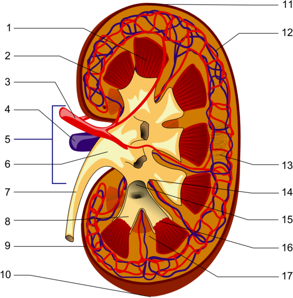
English: Structures of the kidney:
  1. Renal pyramid
  2. Interlobular artery
  3. Renal artery
  4. Renal vein
  5. Renal hylum
  6. Renal pelvis
  7. Ureter
  8. Minor calyx
  9. Renal capsule
  10. Inferior extremity
  11. Superior extremity
  12. Interlobar vein
  13. Nephron
  14. Renal sinus
  15. Major calyx
  16. Renal papilla
  17. Renal column
(no distinction for red/blue (oxygenated or not) blood, arteriole is between capilaries and larger vessels)
日本語: 腎臓の構造:
  1. 腎錐体
  2. 葉間動脈
  3. 腎動脈
  4. 腎静脈
  5. 腎門
  6. 腎盂
  7. 尿管
  8. 小腎杯
  9. 腎被膜
  10. 下端
  11. 上端
  12. 葉間静脈
  13. ネフロン
  14. 腎洞
  15. 大腎杯
  16. 腎乳頭
  17. 腎柱
Español: Partes del riñón:
  1. Pirámide renal
  2. Arteria interlobulillar
  3. Arteria renal
  4. Vena renal
  5. Hilio renal
  6. Pelvis renal
  7. Uréter
  8. Cáliz menor
  9. Cápsula renal
  10. Polo renal inferior
  11. Polo renal superior
  12. Vena interlobulillar
  13. Nefrona
  14. Seno renal
  15. Cáliz mayor
  16. Papila renal
  17. Columna renal
Deutsch: Schema des makroskopischen Aufbaus der Niere:
  1. Nierenmark mit Markkegeln (Pyramides renales)
  2. Vas afferens
  3. Nierenarterie (Arteria renalis)
  4. Nierenvene (Vena renalis)
  5. Hilum renale
  6. Nierenbecken (Pelvis renalis)
  7. Harnleiter (Ureter)
  8. kleine Nierenkelche (Calices minores renales)
  9. Nierenkapsel (Capsula fibrosa renalis)
  10. unterer Nierenpol (Extremitas inferior)
  11. oberer Nierenpol (Extremitas superior)
  12. Vas efferens
  13. Nephron
  14. Nierenbucht (Sinus renalis)
  15. große Nierenkelche (Calices majores renales)
  16. Spitzen der Markkegel (Papillae renales)
  17. Bertini-Säulen (Columna renalis)
Türkçe: Böbreğin yapısı:
  1. Renal piramit (pyramides renales, Malpighi piramitleri)
  2. Interlobar arter (a. interlobaris)
  3. Renal arter (a. renalis)
  4. Renal ven (v. renalis)
  5. Renal hilus (böbrek hilusu, hilum renale)
  6. Renal pelvis (pelvis renalis)
  7. Üreter
  8. Minör kaliks (calices minores renales)
  9. Renal kapsül (capsula fibrosa renalis)
  10. Alt pol (inferior pol, extremitas inferior)
  11. Üst pol (superior pol, extremitas superior)
  12. Interlobar ven (v. interlobaris)
  13. Nefron
  14. Renal sinüs (sinus renalis)
  15. Majör kaliks (calices majores renales)
  16. Renal papilla (papilla renalis)
  17. Renal column (columna renalis, Bertin sütunları)
Polski: Prawa nerka ludzka, przekrój podłużny:
  1. piramida nerkowa
  2. tętnica międzypłatowa
  3. tętnica nerkowa
  4. żyła nerkowa
  5. wnęka nerki
  6. miedniczka nerkowa
  7. moczowód
  8. kielich mniejszy
  9. torebka włóknista
  10. biegun dolny nerki
  11. biegun górny nerki
  12. żyła międzypłatowa
  13. nefron
  14. zatoka nerkowa
  15. kielich większy
  16. brodawka nerkowa
  17. słupy nerkowe
Français : Structure interne d'un rein
  1. Pyramide rénale
  2. Artère interlobaire
  3. Artère rénale
  4. Veine rénale
  5. Hile du rein
  6. Pelvis rénale
  7. Uretère
  8. Calices mineurs
  9. Capsule rénale
  10. Pôle inférieur
  11. Pôle supérieur
  12. Veine interlobaire
  13. Néphron
  14. Sinus rénal
  15. Calices majeurs
  16. Papilles rénales
  17. Colonnes rénales
Русский: Строение почки
  1. Мозговое вещество и почечные пирамиды (Pyramides renales)
  2. Выносящая клубочковая артериола (Arteriola glomerularis efferens)
  3. Почечная артерия (Arteria renalis)
  4. Почечная вена (Vena renalis)
  5. Почечные ворота (Hilus renalis)
  6. Почечная лоханка (Pelvis renalis)
  7. Мочеточник (Ureter)
  8. Малая почечная чашка (Calices minores renales)
  9. Фиброзная капсула почки (Capsula fibrosa renalis)
  10. Нижний полюс почки (Extremitas inferior)
  11. Верхний полюс почки (Extremitas superior)
  12. Приносящая клубочковая артериола (Arteriola glomerularis afferens)
  13. Нефрон (Nephron)
  14. Почечная пазуха (Sinus renalis)
  15. Большая почечная чашка (Calices majores renales)
  16. Вершина почечной пирамиды (Papillae renales)
  17. Почечный столб (Columna renalis)
Italiano: Struttura interna di un rene:
  1. Piramide di Malpighi
  2. Arterie interlobari
  3. Arteria renale
  4. Vena renale
  5. Ileo renale
  6. Pelvi
  7. Uretere
  8. Calice minore
  9. Capsula renale
  10. Estremità inferiore
  11. Estremità superiore
  12. Vene interlobari
  13. Nefrone
  14. Seni renali
  15. Calice maggiore
  16. Papilla renale
  17. Colonna renale
(no distinction for red/blue (oxygenated or not) blood, arteriole is between capilaries and larger vessels)
Date (UTC)
Source Own work
Author Piotr Michał Jaworski; PioM EN DE PL

Place: POLAND/Poznań;
EN: If you want to use my graphic outside Wikipedia, and its resolution or license doesn't satisfy you, write to me: .
PL: Jeżeli chcesz użyć mojej grafiki poza Wikipedią a jej rozdzielczość, bądź licencja nie odpowiadają tobie napisz do mnie: .

Licensing

I, the copyright holder of this work, hereby publish it under the following licenses:
GNU head Permission is granted to copy, distribute and/or modify this document under the terms of the GNU Free Documentation License, Version 1.2 or any later version published by the Free Software Foundation; with no Invariant Sections, no Front-Cover Texts, and no Back-Cover Texts. A copy of the license is included in the section entitled GNU Free Documentation License.
w:en:Creative Commons
attribution share alike
This file is licensed under the Creative Commons Attribution-Share Alike 3.0 Unported license.
You are free:
  • to share – to copy, distribute and transmit the work
  • to remix – to adapt the work
Under the following conditions:
  • attribution – You must give appropriate credit, provide a link to the license, and indicate if changes were made. You may do so in any reasonable manner, but not in any way that suggests the licensor endorses you or your use.
  • share alike – If you remix, transform, or build upon the material, you must distribute your contributions under the same or compatible license as the original.
This licensing tag was added to this file as part of the GFDL licensing update.
You may select the license of your choice.

Wikipedia

 This is a featured picture on the Polish language Wikipedia (Ilustracja na medal) and is considered one of the finest images. See its nomination here.
 This is a featured picture on the Turkish language Wikipedia (Seçkin resimler) and is considered one of the finest images. See its nomination here.

If you think this file should be featured on Wikimedia Commons as well, feel free to nominate it.
If you have an image of similar quality that can be published under a suitable copyright license, be sure to upload it, tag it, and nominate it.

Captions

Add a one-line explanation of what this file represents

Items portrayed in this file

depicts

8 June 2006

File history

Click on a date/time to view the file as it appeared at that time.

Date/TimeThumbnailDimensionsUserComment
current10:06, 10 June 2006Thumbnail for version as of 10:06, 10 June 20061,000 × 1,018 (343 KB)Piomlittle changes
07:37, 10 June 2006Thumbnail for version as of 07:37, 10 June 20061,000 × 1,018 (342 KB)Piomsome little changes
07:23, 10 June 2006Thumbnail for version as of 07:23, 10 June 20061,000 × 1,018 (342 KB)Piomtraditional numbering
20:57, 9 June 2006Thumbnail for version as of 20:57, 9 June 20061,000 × 983 (401 KB)Piomnowe piramidki dla WarXa
18:34, 9 June 2006Thumbnail for version as of 18:34, 9 June 20061,000 × 983 (495 KB)Piomsome changes in shapes
19:54, 8 June 2006Thumbnail for version as of 19:54, 8 June 2006800 × 701 (278 KB)Piomcorected numbers
19:48, 8 June 2006Thumbnail for version as of 19:48, 8 June 2006800 × 701 (361 KB)Piom{{subst:User:Piom/a|~~~~~|Kidney anatomy|Human_anatomy}}

Global file usage

The following other wikis use this file:

View more global usage of this file.

Metadata